Pes Anserinus Ultrasound Images !!
pes anserinus tendons browse free ultrasound cases ultrasound.
ultrasound images clips pes anserinus tendinitis and bursitis pes anserinus tendinitis with thickened tendons and fluid filled bursa 2014 he was the head.
pes anserinus radiology reference article radiopaedia org.
the pes anserinus inserts on the medial side of the tibial tuberosity below or distal to the tibial tuberosity with significant variant anatomy 3 4 comprising mostly different accessory tendinous bands appearing from the different tendons the type of insertion can be classified into a short band shaped and fan shaped with fan shaped defined.pes anserinus tendons browse free ultrasound cases ultrasound.
06 06 2013 ultrasound images clips pes anserinus tendinitis with thickened pes anserinus tendons and peritendinous effusion and a fluid filled bursa he was the head of the.
pes anserinus tendons browse free ultrasound cases ultrasound.
06 06 2013 ultrasound images clips pes anserinus tendinitis and bursitis with peritendinous effusion and a fluid filled bursa the effusion also extends around the semimembranosus tendon.pes anserinus tendons browse free ultrasound cases ultrasound.
dr taco geertsma is the founder of ultrasoundcases info and a retired radiologist and has worked in the gelderse vallei hospital from january 1 1983 till july 1 2014 he was the head of the ultrasound department for many years.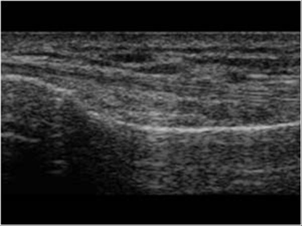
knee ultrasound radiology key.
06 03 2019 here the pes anserinus can be seen as three hyperechoic tendons superficial to the tibial collateral ligament that converge onto the tibia toggling the transducer is often helpful because this will cause the tendons of the pes anserinus superficial to the tibial collateral ligament to appear hypoechoic from anisotropy and be more conspicuous.pes anserinus bursitis radiology reference article.
pes anserinus bursitis refers to symptomatic inflammation of the pes anserinus bursa which is located at the medial aspect of the knee at the level of the joint space deep to the pes anserinus tendons clinical presentation classically symp.
pes anserine tendinopathy bursitis ultrasound guided injections.
the pes anserine tendons attach three muscles from the inside of the thigh onto a common insertion point on the inside of the tibia bone.ultrasound pes anserine bursa exam the pain source makes.
03 12 2010 by chris faubel m d bertolotti s syndrome is an atypical cause of axial low back pain or buttock pain caused by a transitional lumbar vertebrae with a large transverse process that either fuses with the sacrum sacral ala or ilium or forms a pseudoarticulation at that location.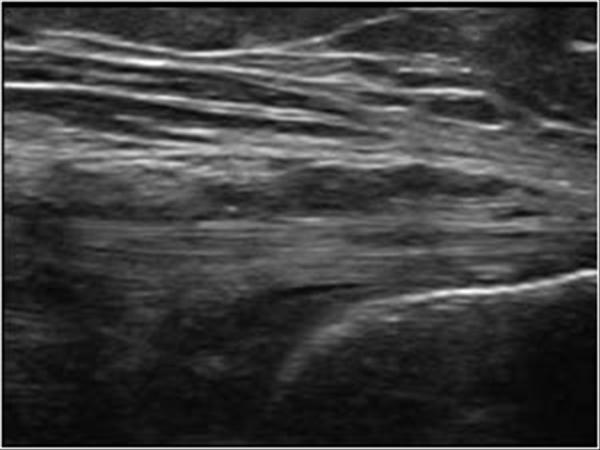
pes anserinus ultrasound images
pes anserinus ultrasound images
pes adalah,pes anserinus,pes andri,pes android,pes apk,pes anserine bursitis,pes adalah penyakit,pes anserinus tendinitis,pes anserinus adalah,pes anserine,anserinus ab,astragalus anserinus,pes anserinus anatomy,pes anserinus attachment site,pes anserinus action,pes anserinus attachment,pes anserinus arrangement is found in which nerve,pes anserine area,pes anserinus acl reconstruction,pes anserinus anatomy picture,ultrasound adalah,ultrasound assisted extraction,ultrasound abdomen,ultrasound assisted extraction adalah,ultrasound and infrasound,ultrasound adalah pdf,ultrasound at 6 weeks,ultrasound at 5 weeks,ultrasound at 8 weeks,ultrasound abdomen and pelvis,images artinya,images and words,images adalah,images aloe vera gel,images aloe vera gel review,images api,images assalamualaikum warahmatullahi wabarakatuh,images are formed by eye lens at,images and shades,images app
Posting Komentar untuk "Pes Anserinus Ultrasound Images !!"